Peel không bong – Xu hướng tất yếu của ngành làm đẹp an toàn Ngành làm đẹp đang bị siết chặt quản lý với...
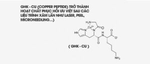
Trong kỷ nguyên công nghệ sinh học lên ngôi, Copper Peptide – cụ thể là GHK-Cu (Glycyl-L-Histidyl-L-Lysine Copper Complex) – đã được khẳng định vai trò như một hoạt chất sinh học đa tác động.
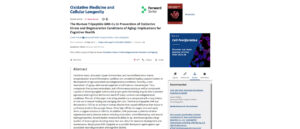
Từ thập niên 1970, nhiều công trình nghiên cứu, nổi bật là báo cáo đăng trên Oxidative Medicine and Cellular Longevity (Pickart và cộng sự, 2012), đã chỉ ra rằng GHK-Cu không chỉ phục hồi mô tổn thương, kích thích tăng sinh collagen, mà còn điều hòa gen, chống oxy hóa và kháng viêm mạnh mẽ – những cơ chế then chốt để duy trì sự trẻ trung của làn da và sức khỏe tế bào.
Dựa trên nền tảng khoa học đó, Merislab Pure Copper Peptide Ampoule ra đời, ứng dụng công thức độc quyền Copper Peptide Complex®, kết hợp GHK-Cu cùng các thành phần sinh học tối ưu. Đây được xem là giải pháp thế hệ mới cho làn da tổn thương, nhạy cảm và đang bước vào giai đoạn lão hóa.
Sức mạnh chống oxy hóa: Bảo vệ cấu trúc tế bào và DNA khỏi stress oxy hóa
Cơ chế lão hóa da khởi phát từ sự tích tụ các gốc tự do (Reactive Oxygen Species – ROS). Chúng phá hủy lipid màng tế bào, biến đổi protein và làm tổn thương DNA – hệ quả là mất cân bằng nội môi, hình thành nếp nhăn, giảm độ đàn hồi.
GHK-Cu đã được chứng minh trong các nghiên cứu in vitro và in vivo có khả năng:
> Tăng hoạt tính Superoxide Dismutase (SOD1) – enzyme chủ lực vô hiệu hóa gốc tự do (Pickart, 2012).
> Giảm đáng kể quá trình peroxid hóa lipid, bảo vệ màng tế bào và protein cấu trúc.
> Ngăn chặn tích tụ ion sắt tự do, vốn là chất xúc tác gây tổn thương oxy hóa nặng nề trong da và mô (Pickart, 1985).
Tính năng chống oxy hóa đặc biệt này giúp GHK-Cu bảo vệ toàn diện cấu trúc nền da, làm chậm quá trình lão hóa từ gốc.
Kháng viêm và điều hòa miễn dịch: Hạn chế tăng sắc tố và thoái hóa mô
Viêm mạn tính mức độ thấp (inflammaging) là yếu tố trung tâm thúc đẩy lão hóa và tăng sắc tố sau viêm (PIH). Các cytokine như TNF-alpha, TGF-beta không chỉ kích hoạt melanocyte mà còn làm suy giảm chức năng nguyên bào sợi.
Nghiên cứu của McCormack (2001) và Canapp (2003) chỉ ra rằng GHK-Cu có khả năng ức chế rõ rệt TNF-alpha, TGF-beta, từ đó:
> Giảm viêm sưng cấp tính và mạn tính
> Ngăn ngừa hình thành sẹo xơ và rối loạn sắc tố
> Duy trì môi trường vi mô lý tưởng cho tái tạo mô
Nhờ cơ chế điều hòa miễn dịch thông minh, Copper Peptide trở thành hoạt chất phục hồi ưu việt sau các liệu trình xâm lấn như laser, peel, microneedling…
Thúc đẩy tăng sinh collagen, elastin và tái tạo mạch máu
Một trong những ưu điểm nổi bật nhất của GHK-Cu là khả năng kích thích tổng hợp collagen, elastin và glycosaminoglycan, trực tiếp cải thiện độ dày, độ săn chắc và đàn hồi của da:
> Maquart (1988–2000) chứng minh nồng độ GHK-Cu chỉ 1–10 nanomolar đã đủ để:
- Tăng tổng hợp collagen type I và III.
- Kích thích sản xuất decorin – proteoglycan điều hòa cấu trúc collagen.
- Cân bằng hoạt tính metalloproteinase (MMPs) và ức chế TIMPs – quá trình tái cấu trúc collagen tối ưu.
> Sage (1991) phát hiện GHK còn kích thích biểu hiện VEGF, FGF, hỗ trợ tái tạo vi tuần hoàn và phục hồi độ hồng hào tự nhiên của da.
Đây chính là nền tảng giúp Copper Peptide phục hồi toàn diện mô da sau tổn thương và nâng cấp chất lượng da lão hóa.
Công nghệ Copper Peptide Complex® trong Merislab Pure Copper Peptide Ampoule
Merislab Pure Copper Peptide Ampoule là sản phẩm thế hệ mới ứng dụng công nghệ Copper Peptide Complex®, một nền tảng công nghệ độc quyền kết hợp Copper Peptide tinh khiết cùng các thành phần hỗ trợ sinh học tối ưu như EGF, Sodium Hyaluronic Acid và chiết xuất nấm tuyết.
Sự kết hợp này mang đến tác động hiệp lực đa chiều: thúc đẩy tốc độ sản xuất collagen và elastin, làm lành nhanh các vi tổn thương sau liệu trình xâm lấn, đồng thời ngăn chặn hình thành tăng sắc tố hậu viêm – một trong những vấn đề nan giải của da nhạy cảm hoặc da đang tái tạo.
Bên cạnh đó, Copper Peptide Complex® còn phát huy hiệu quả điều hòa hoạt động gen trong tế bào da, giúp khôi phục khả năng tự tái tạo, sửa chữa DNA và tăng sinh các yếu tố tăng trưởng nội sinh, từ đó cải thiện rõ rệt độ dày, độ săn chắc, độ đàn hồi của lớp nền da.
Lợi ích nổi bật được chứng minh:
> Phục hồi tổn thương và ngăn sạm sau viêm
> Thúc đẩy tái tạo collagen – elastin
> Giảm nếp nhăn, trẻ hóa bề mặt da
> Cải thiện khả năng chống oxy hóa nội sinh
> Tăng hiệu quả các sản phẩm treatment
> Tăng tốc lành thương sau peel, laser, microneedling
Từ dữ liệu khoa học uy tín suốt nhiều thập kỷ, Copper Peptide (GHK-Cu) đã được khẳng định là hoạt chất đa nhiệm bậc nhất trong ngành thẩm mỹ: chống oxy hóa, kháng viêm, kích thích tổng hợp collagen và phục hồi chức năng tế bào.
Sự ra đời của Merislab Pure Copper Peptide Ampoule không chỉ tiếp nối di sản nghiên cứu, mà còn nâng cấp Copper Peptide lên tầm cao mới nhờ công nghệ kết hợp tối ưu, đáp ứng nhu cầu chăm sóc da chuyên sâu, an toàn và bền vững.
Nếu bạn đang tìm kiếm giải pháp phục hồi – trẻ hóa – tái sinh toàn diện, Copper Peptide chính là chìa khóa vàng giúp làn da lấy lại sự khỏe mạnh, căng bóng và trẻ trung lâu dài.
—
TÀI LIỆU THAM KHẢO
1 Shukla V., Mishra S. K., and Pant H. C., Oxidative stress in neurodegeneration, Advances Pharmacological Sciences. (2011) 2011, 13, 572634, https://doi.org/10.1155/2011/572634.
View
Google Scholar
2 McGeer P. L. and McGeer E. G., Inflammation and the degenerative diseases of aging, Annals of the New York Academy of Sciences. (2004) 1035, 104–116, 2-s2.0-14944352071, https://doi.org/10.1196/annals.1332.007.
Web of Science®
3 Iraola-Guzmán S., Estivill X., and Rabionet R., DNA methylation in neurodegenerative disorders: a missing link between genome and environment?, Clinical Genetics. (2011) 80, no. 1, 1–14, 2-s2.0-79958170373, https://doi.org/10.1111/j.1399-0004.2011.01673.x.
4 Moosmann B. and Behl C., Antioxidants as treatment for neurodegenerative disorders, Expert Opinion on Investigational Drugs. (2002) 11, no. 10, 1407–1435, 2-s2.0-0036799799, https://doi.org/10.1517/13543784.11.10.1407.
Web of Science®
5 Kamat C. D., Gadal S., Mhatre M., Williamson K. S., Pye Q. N., and Hensley K., Antioxidants in central nervous system diseases: preclinical promise and translational challenges, Journal of Alzheimer′s Disease. (2008) 15, no. 3, 473–493, 2-s2.0-57349180534.
Web of Science®
6 Jomova K. and Valko M., Importance of iron chelation in free radical-induced oxidative stress and human disease, Current Pharmaceutical Design. (2011) 17, no. 31, 3460–3473.
7 Kwok J. B., Role of epigenetics in Alzheimer′s and Parkinson′s disease, Epigenomics. (2010) 2, no. 5, 671–682, 2-s2.0-78149433116, https://doi.org/10.2217/epi.10.43.
8 Pickart L., The human tri-peptide GHK and tissue remodeling, Journal of Biomaterials Science, Polymer Edition. (2008) 19, no. 8, 969–988, 2-s2.0-46849097170, https://doi.org/10.1163/156856208784909435.
Web of Science®
9 Pickart L. and Pickart F., A possible mechanism whereby skin remodeling may suppress cancer metastasis genes, Society for the Advancement of Wound Care and the Wound Healing Society, 2011, Wound Repair and Regeneration, Dallas, Tex, USA, A8–A62.
10 Pickart L., A tripepeptide in human serum that promotes the growth of hepatoma cells and the survival of normal hepatocytes, Ph.D. thesis, 1973, University of California, San Francisco, Calif, USA.
11 Pickart L., Freedman J. H., and Loker W. J., Growth-modulating plasma tripeptide may function by facilating copper uptake into cells, Nature. (1980) 288, no. 5792, 715–717, 2-s2.0-0019155815.
12 Perkins C. M., Rose N. J., Weinstein B., Stenkamp R. E., Jensen L. H., and Pickart L., The structure of a copper complex of the growth factor glycyl-L-histidyl-L-lysine at 1.1 Å resolution, Inorganica Chimica Acta. (1984) 82, no. 1, 93–99, 2-s2.0-0006186651.
13 Hureau C., Eury H., Guillot R., Bijani C., Sayen S., Solari P. L., Guillon E., Faller P., and Dorlet P., X-ray and solution structures of Cu(II) GHK and Cu(II) DAHK complexes: influence on their redox properties, Chemistry A. (2011) 17, 10151–10160.
14 Lau S. J. and Sarkar B., The interaction of copper(II) and glycyl-L-histidyl-L-lysine, a growth-modulating tripeptide from plasma, Biochemical Journal. (1981) 199, no. 3, 649–656, 2-s2.0-0019834173.
15 Kumar N., GrossJ. B.Jr., and Ahlskog J. E., Copper deficiency myelopathy produces a clinical picture like subacute combined degeneration, Neurology. (2004) 63, no. 1, 33–39, 2-s2.0-3242718536.
16 Kazemi A., Frazier T., and Cave M., Micronutrient-related neurologic complications following bariatric surgery, Current Gastroenterology Reports. (2010) 12, no. 4, 288–295, 2-s2.0-77956267962, https://doi.org/10.1007/s11894-010-0120-5.
17 Naismith R. T., Shepherd J. B., Weihl C. C., Tutlam N. T., and Cross A. H., Acute and bilateral blindness due to optic neuropathy associated with copper deficiency, Archives of Neurology. (2009) 66, no. 8, 1025–1027, 2-s2.0-68549135352, https://doi.org/10.1001/archneurol.2009.70.
18 Zucconi G. G., Cipriani S., Scattoni R., Balgkouranidou I., Hawkins D. P., and Ragnarsdottir K. V., Copper deficiency elicits glial and neuronal response typical of neurodegenerative disorders, Neuropathology and Applied Neurobiology. (2007) 33, no. 2, 212–225, 2-s2.0-33847759150, https://doi.org/10.1111/j.1365-2990.2006.00793.x.
19 Hung Y. H., Bush A. I., and Cherny R. A., Copper in the brain and Alzheimer′s disease, Journal of Biological Inorganic Chemistry. (2010) 15, no. 1, 61–76, 2-s2.0-72049102879, https://doi.org/10.1007/s00775-009-0600-y.
20 Bolognin S., Messori L., Drago D., Gabbiani C., Cendron L., and Zatta P., Aluminum, copper, iron and zinc differentially alter amyloid-Aβ (1–42) aggregation and toxicity, International Journal of Biochemistry and Cell Biology. (2011) 43, no. 6, 877–885, 2-s2.0-79955927181, https://doi.org/10.1016/j.biocel.2011.02.009.
21 Exley C., Aluminium and iron, but neither copper nor zinc, are key to the precipitation of β-sheets of Aβ42 in senile plaque cores in Alzheimer′s disease, Journal of Alzheimer′s Disease. (2006) 10, no. 2-3, 173–177, 2-s2.0-33751107948.
22 Kawahara M., Koyama H., Nagata T., and Sadakane Y., Zinc, copper, and carnosine attenuate neurotoxicity of prion fragment PrP106–126, Metallomics. (2011) 3, no. 7, 726–734, 2-s2.0-79960152379, https://doi.org/10.1039/c1mt00015b.
23 Bishop G. M. and Robinson S. R., The amyloid paradox: Amyloid-β-metal complexes be neurotoxic and neuroprotective, Brain Pathology. (2004) 14, no. 4, 448–452, 2-s2.0-8744255638.
24 Schrag M., Mueller C., Oyoyo U., Smith M. A., and Kirsch W. M., Iron, zinc and copper in the Alzheimer′s disease brain: a quantitative meta-analysis. Some insight on the influence of citation bias on scientific opinion, Progress in Neurobiology. (2011) 94, no. 3, 296–306, 2-s2.0-79959395494, https://doi.org/10.1016/j.pneurobio.2011.05.001.
Web of Science®
25 Kessler H., Pajonk F. G., Meisser P., Schneider-Axmann T., Hoffmann K. H., Supprian T., Herrmann W., Obeid R., Multhaup G., Falkai P., and Bayer T. A., Cerebrospinal fluid diagnostic markers correlate with lower plasma copper and ceruloplasmin in patients with Alzheimer′s disease, Journal of Neural Transmission. (2006) 113, no. 11, 1763–1769, 2-s2.0-33750463720, https://doi.org/10.1007/s00702-006-0485-7.
26 Doraiswamy P. M. and Finefrock A. E., Metals in our minds: therapeutic implications for neurodegenerative disorders, Lancet Neurology. (2004) 3, no. 7, 431–434, 2-s2.0-2942729775, https://doi.org/10.1016/S1474-4422(04)00809-9.
27 Akatsu H., Hori A., Yamamoto T. et al., Transition metal abnormalities in progressive dementias, BioMetals. (2012) 52, no. 2, 337–350, https://doi.org/10.1007/s10534-011-9504-8.
28 Kessler H., Pajonk F. G., Bach D., Schneider-Axmann T., Falkai P., Herrmann W., Multhaup G., Wiltfang J., Schäfer S., Wirths O., and Bayer T. A., Effect of copper intake on CSF parameters in patients with mild Alzheimer′s disease: a pilot phase 2 clinical trial, Journal of Neural Transmission. (2008) 115, no. 12, 1651–1659, 2-s2.0-57049180732, https://doi.org/10.1007/s00702-008-0136-2.
29 Borchardt T., Camakaris J., Cappai R., Masters C. L., Beyreuther K., and Multhaup G., Copper inhibits β-amyloid production and stimulates the non-amyloidogenic pathway of amyloid-precursor-protein secretion, Biochemical Journal. (1999) 344, 461–467, 2-s2.0-0033430087, https://doi.org/10.1042/0264-6021:3440461.
30 Narahara M., Hamada-Kanazawa M., Kouda M., Odani A., and Miyake M., Superoxide scavenging and xanthine oxidase inhibiting activities of copper-β-citryl-L-glutamate complex, Biological and Pharmaceutical Bulletin. (2010) 33, no. 12, 1938–1943, 2-s2.0-78751481404, https://doi.org/10.1248/bpb.33.1938.
31 Rózga M. and Bal W., The Cu(II)/Aβ/human serum albumin model of control mechanism for copper-related amyloid neurotoxicity, Chemical Research in Toxicology. (2010) 23, no. 2, 298–308, 2-s2.0-77049095207, https://doi.org/10.1021/tx900358j.
32 Perrone L., Mothes E., Vignes M., Mockel A., Figueroa C., Miquel M. C., Maddelein M. L., and Faller P., Copper transfer from Cu-Aβ to human serum albumin inhibits aggregation, radical production and reduces Aβ toxicity, ChemBioChem. (2010) 11, no. 1, 110–118, 2-s2.0-75649136954, https://doi.org/10.1002/cbic.200900474.
33 Lutsenko S., Bhattacharjee A., and Hubbard A. L., Copper handling machinery of the brain, Metallomics. (2010) 2, no. 9, 596–608, 2-s2.0-77956330693, https://doi.org/10.1039/c0mt00006j.
34 Hawk S. N., Lanoue L., Keen C. L., Kwik-Uribe C. L., Rucker R. B., and Uriu-Adams J. Y., Copper-deficient rat embryos are characterized by low superoxide dismutase activity and elevated superoxide anions, Biology of Reproduction. (2003) 68, no. 3, 896–903, 2-s2.0-0037369760, https://doi.org/10.1095/biolreprod.102.009167.
35 Bayer T. A., Schäfer S., Simons A., Kemmling A., Kamer T., Tepest R., Eckert A., Schüssel K., Eikenberg O., Sturchler-Pierrat C., Abramowski D., Staufenbiel M., and Multhaup G., Dietary Cu stabilizes brain superoxide dismutase 1 activity and reduces amyloid Aβ production in APP23 transgenic mice, Proceedings of the National Academy of Sciences of the United States of America. (2003) 100, no. 24, 14187–14192, 2-s2.0-10744226316, https://doi.org/10.1073/pnas.2332818100.
36 Arul V., Gopinath D., Gomathi K., and Jayakumar R., GHK peptide incorporated collagenous matrix: a novel biomaterial for dermal wound healing in rats, Journal of Biomedical Materials Research. (2005) 73, no. 2, 383–391.
37 Miller D. M., DeSilva D., Pickart L., and Aust S. D., Effects of glycyl-histidyl-lysyl chelated Cu(II) on ferritin dependent lipid peroxidation, Advances in Experimental Medicine and Biology. (1990) 264, 79–84, 2-s2.0-0025180049.
38 Alberghina M., Lupo G., La Spina G., Mangiameli A., Gulisano M., Sciotto D., and Rizzarelli E., Cytoprotective effect of copper(II) complexes against ethanol-induced damage to rat gastric mucosa, Journal of Inorganic Biochemistry. (1992) 45, no. 4, 245–259, 2-s2.0-0026582790, https://doi.org/10.1016/0162-0134(92)84013-D.
CAS
PubMed
Web of Science®
39 Beretta G., Artali R., Regazzoni L., Panigati M., and Facino R. M., Glycyl-histidyl-lysine (GHK) is a quencher of α,β-4-hydroxy-trans-2-nonenal: a comparison with carnosine. Insights into the mechanism of reaction by electrospray ionization mass spectrometry, 1H NMR, and computational techniques, Chemical Research in Toxicology. (2007) 20, no. 9, 1309–1314, 2-s2.0-35148824739, https://doi.org/10.1021/tx700185s.
40 Beretta G., Arlandini E., Artali R., Anton J. M., and Maffei Facino R., Acrolein sequestering ability of the endogenous tripeptide glycyl-histidyl-lysine (GHK): characterization of conjugation products by ESI-MSn and theoretical calculations, Journal of Pharmaceutical and Biomedical Analysis. (2008) 47, no. 3, 596–602, 2-s2.0-44249095307, https://doi.org/10.1016/j.jpba.2008.02.012.
41 Agostinho P., Cunha R. A., and Oliveira C., Neuroinflammation, oxidative stress and the pathogenesis of Alzheimer′s disease, Current Pharmaceutical Design. (2010) 16, no. 25, 2766–2778, 2-s2.0-77957970155.
42 McCormack M. C., Nowak K. C., and Koch R. J., The effect of copper tripeptide and tretinoin on growth factor production in a serum-free fibroblast model., Archives of Facial Plastic Surgery. (2001) 3, no. 1, 28–32, 2-s2.0-0035229667.
43 CanappS. O.Jr., Farese J. P., Schultz G. S., Gowda S., Ishak A. M., Swaim S. F., Vangilder J., Lee-Ambrose L., and Martin F. G., The effect of topical tripeptide-copper complex on healing of ischemic open wounds, Veterinary Surgery. (2003) 32, no. 6, 515–523, 2-s2.0-1542603606, https://doi.org/10.1111/j.1532-950X.2003.00515.x.
44 Kalaria R. N., Vascular basis for brain degeneration: Faltering controls and risk factors for dementia, Nutrition Reviews. (2010) 68, no. supplement 2, S74–S87, 2-s2.0-78649427539, https://doi.org/10.1111/j.1753-4887.2010.00352.x.
45 Pollard J. D., Quan S., Kang T., and Koch R. J., Effects of copper tripeptide on the growth and expression of growth factors by normal and irradiated fibroblasts, Archives of Facial Plastic Surgery. (2005) 7, no. 1, 27–31, 2-s2.0-17144407587, https://doi.org/10.1001/archfaci.7.1.27.
46 Lane T. F., Iruela-Arispe M. L., Johnson R. S., and Sage E. H., SPARC is a source of copper-binding peptides that stimulate angiogenesis, Journal of Cell Biology. (1994) 125, no. 4, 929–943, 2-s2.0-0028355889, https://doi.org/10.1083/jcb.125.4.929.
47 Numakawa T., Matsumoto T., Numakawa Y. et al., Protective action of neurotrophic factors and estrogen against oxidative stress-mediated neurodegeneration, Journal of Toxicology. (2011) 2011, 12, 405194, https://doi.org/10.1155/2011/405194.
48 Sensenbrenner M., Jaros G. G., Moonen G., and Meyer B. J., Effect of conditioned media on nerve cell differentiation, Experientia. (1980) 36, no. 6, 660–662, 2-s2.0-0019152047.
49 Lindner G., Grosse G., Halle W., and Henklein P., The effect of a synthetic tripeptide nervous tissue cultured in vitro, Zeitschrift für Mikroskopisch-Anatomische Forschung. (1979) 93, no. 5, 820–828.
50 Ahmed M. R., Basha S. H., Gopinath D., Muthusamy R., and Jayakumar R., Initial upregulation of growth factors and inflammatory mediators during nerve regeneration in the presence of cell adhesive peptide-incorporated collagen tubes, Journal of the Peripheral Nervous System. (2005) 10, no. 1, 17–30, 2-s2.0-14244252868, https://doi.org/10.1111/j.1085-9489.2005.10105.x.
51 Spencer J. P., Vafeiadou K., Williams R. J., and Vauzour D., Neuroinflammation: modulation by flavonoids and mechanisms of action, Molecular Aspects of Medicine. (2012) 33, no. 1, 83–97.
52 Wang J. Y., Wen L. L., Huang Y. N., Chen Y. T., and Ku M. C., Dual effects of antioxidants in neurodegeneration: direct neuroprotection against oxidative stress and indirect protection via suppression of glia-mediated inflammation, Current Pharmaceutical Design. (2006) 12, no. 27, 3521–3533, 2-s2.0-33748750998, https://doi.org/10.2174/138161206778343109.
53 Pickart L., Use of GHL-Cu as a wound-healing and anti-inflammatory agent, United States Patent 4,760,051, July 1988.
54 Downey D., Larrabee W. F., Voci V., and Pickart L., Acceleration of wound healing using glycyl-histidyl-lysyl Cu(II), Surgical Forum. (1985) 36, 573–575, 2-s2.0-0022406029.
55 Maquart F. X., Pickart L., Laurent M., Gillery P., Monboisse J. C., and Borel J. P., Stimulation of collagen synthesis in fibroblast cultures by the tripeptide-copper complex glycl-L-histidyl-L-lysine-Cu2+, FEBS Letters. (1988) 238, no. 2, 343–346, 2-s2.0-0023719508.
56 Wegrowski Y., Maquart F. X., and Borel J. P., Stimulation of sulfated glycosaminoglycan synthesis by the tripeptide-copper complex glycyl-L-histidyl-L-lysine-Cu2+, Life Sciences. (1992) 51, no. 13, 1049–1056, 2-s2.0-0026757171.
57 Siméon A., Emonard H., Hornebeck W., and Maquart F. X., The tripeptide-copper complex glycyl-L-histidyl-L-lysine-Cu2+ stimulates matrix metalloproteinase-2 expression by fibroblast cultures, Life Sciences. (2000) 67, no. 18, 2257–2265, 2-s2.0-0034703502, https://doi.org/10.1016/S0024-3205(00)00803-1.
58 Siméon A., Wegrowski Y., Bontemps Y., and Maquart F. X., Expression of glycosaminoglycans and small proteoglycans in wounds: modulation by the tripeptide-copper complex glycyl-L-histidyl-L-lysine-Cu2+, Journal of Investigative Dermatology. (2000) 115, no. 6, 962–968, 2-s2.0-0034521219, https://doi.org/10.1046/j.1523-1747.2000.00166.x.
59 Kang Y. A., Choi H. R., Na J. I., Huh C. H., Kim M. J., Youn S. W., Kim K. H., and Park K. C., Copper-GHK increases integrin expression and p63 positivity by keratinocytes, Archives of Dermatological Research. (2009) 301, no. 4, 301–306, 2-s2.0-67349137554, https://doi.org/10.1007/s00403-009-0942-x.
60 Lamb J., The Connectivity Map: a new tool for biomedical research, Nature Reviews Cancer. (2007) 7, no. 1, 54–60, 2-s2.0-33845876254, https://doi.org/10.1038/nrc2044.
Web of Science®
61 Iorio F., Bosotti R., Scacheri E., Belcastro V., Mithbaokar P., Ferriero R., Murino L., Tagliaferri R., Brunetti-Pierri N., Isacchi A., and Di Bernardo D., Discovery of drug mode of action and drug repositioning from transcriptional responses, Proceedings of the National Academy of Sciences of the United States of America. (2010) 107, no. 33, 14621–14626, 2-s2.0-77957044703, https://doi.org/10.1073/pnas.1000138107.
62 Hong Y., Downey T., Eu K. W., Koh P. K., and Cheah P. Y., A ‘metastasis-prone’ signature for early-stage mismatch-repair proficient sporadic colorectal cancer patients and its implications for possible therapeutics, Clinical and Experimental Metastasis. (2010) 27, no. 2, 83–90, 2-s2.0-77949907451, https://doi.org/10.1007/s10585-010-9305-4.
63 Huang P., Kishida S., Cao D., Murakami-Tonami Y., Mu P., Nakaguro M., Koide N., Takeuchi I., Onishi A., and Kadomatsu K., The neuronal differentiation factor neuroD1 downregulates the neuronal repellent factor Slit2 expression and promotes cell motility and tumor formation of neuroblastoma, Cancer Research. (2011) 71, no. 8, 2938–2948, 2-s2.0-79954591415, https://doi.org/10.1158/0008-5472.CAN-10-3524.
64 Koch C. M. and Wagner W., Epigenetic-aging-signature to determine age in different tissues, Aging. (2011) 3, no. 10, 1018–1027.
65 Fischer A., Sananbenesi F., Mungenast A., and Tsai L. H., Targeting the correct HDAC(s) to treat cognitive disorders, Trends in Pharmacological Sciences. (2010) 31, no. 12, 605–617, 2-s2.0-78449288141, https://doi.org/10.1016/j.tips.2010.09.003.
Web of Science®
66 Hostynek J. J., Dreher F., and Maibach H. I., Human skin retention and penetration of a copper tripeptide in vitro as function of skin layer towards anti-inflammatory therapy, Inflammation Research. (2010) 59, no. 11, 983–988, 2-s2.0-78651279517, https://doi.org/10.1007/s00011-010-0214-4.
67 Hostynek J. J., Dreher F., and Maibach H. I., Human skin penetration of a copper tripeptide in vitro as a function of skin layer, Inflammation Research. (2011) 60, no. 1, 79–86, 2-s2.0-78651366293, https://doi.org/10.1007/s00011-010-0238-9.
Google Scholar
68 Pickart L., Method of using copper(II) containing compounds to accelerate wound healing, United States Patent 5,164,367, November 1992.
Tin tức liên quan
Năm 2024, ngành làm đẹp tại Việt Nam chứng kiến những thay đổi lớn trong chính sách quản lý. Các dịch vụ xâm lấn...
Trong những năm gần đây, xu hướng peel da không bong đã trở thành giải pháp thẩm mỹ an toàn, hiệu quả, được nhiều...